Diagnostic Imaging

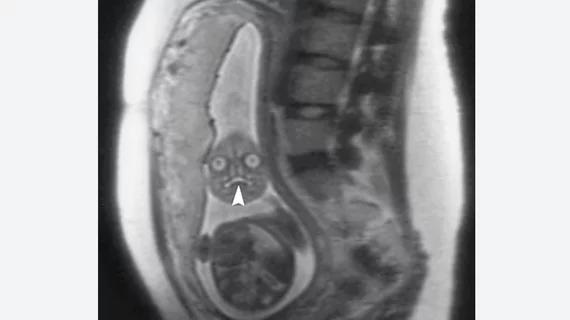
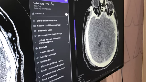
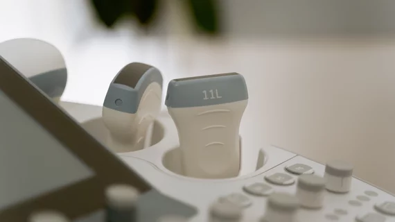
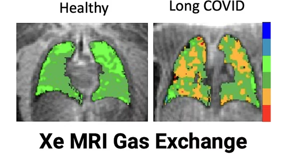
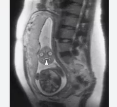
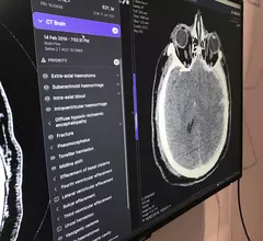
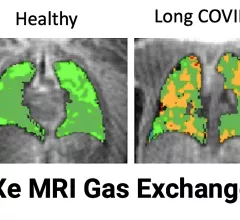
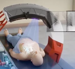
Radiologists use diagnostic imaging to non-invasively look inside the body to help determine the causes of an injury or an illness, and confirm a diagnosis. Providers use many imaging modalities to do so, including CT, MRI, X-ray, Ultrasound, PET and more.

An MRI of a fetus inside the mother. The freakish appearance of the eyes and the face are normal for MRI fetal imaging. Image courtesy of RSNA. Baby MRI, baby ultrasound

PHOTO GALLERY: What does fetal medical imaging look like?

This is a clinical photo gallery of fetal imaging that explains what all can be seen on medical imaging, how sex is determined, how measurements are used to track the development of a baby.

March 11, 2024
Example of data generated by an automated artificial intelligence (AI) brain CT assessment tool from Annalise.ai at RSNA 2022. What does brain imaging look like?

AI company racks up 7 new FDA clearances for image triage and notification solutions

The Australia-based company made the announcement on April 12 in a release that described the timing of these AI-assisted solutions as “increasingly important” amid growing workloads and staffing shortages. 

April 12, 2023

6 pointers on POCUS leadership in the ED (and potentially beyond)

Has point-of-care ultrasound outpaced hospitals’ capacity to incorporate the technology without anointing any particular specialty its proper guardian? The case could be made.

January 28, 2023
Sean Fain, PhD, vice chair of radiology and research and a professor of radiology, Division of Cardiovascular and Thoracic Imaging, University of Iowa, discusses how long-COVID lung damage can be tracked using xenon (Xe) gas magnetic resonance imaging (MRI) and quantitative computed tomography (CT). He spoke to Health Imaging at RSNA 2022.

VIDEO: Tracking long-COVID lung damage using MRI and CT

Sean Fain, PhD, vice chair of radiology and research and a professor of radiology, University of Iowa, discusses how long-COVID lung damage can be tracked using xenon (Xe) gas MRI and quantitative CT at RSNA 2022. 

December 16, 2022
A stack deep silicon photon counting CT detector components. The University of Wisconsin is working with GE Healthcare to develop the new PCD technology. The system and the deep silicon technology was unveiled at RSNA 2022. #RSNA22 #PCCT

VIDEO: Photon-counting CT development at the University of Wisconsin

Tim Szczykutowicz, PhD, DABR, associate professor of radiology at the University of Wisconsin-Madison, is helping develop a new type of photon-counting CT detector that was shown as a work-in-progress by GE Healthcare at RSNA 2022.

December 13, 2022
Bruce Haffty, MD, president of Radiological Society of North America (RSNA) Board of Directors, chair of radiation oncology, associate vice chancellor for cancer programs at Rutgers Cancer Institute of New Jersey and system director of radiation oncology at RWJBarnabas Health, discusses the president’s address "Diagnostic Imaging: Value From the Lens of the Patient," with Radiology Business. Haffty will present his opening session at RSNA 2022 at 4 p.m., Nov. 27, 2022, in the Arie Crown theater.  

VIDEO: RSNA President Bruce Haffty previews the big conference in Chicago

Haffty offered his perspective on the conference as a whole and previewed his presidential address, which is scheduled for early Sunday evening.

November 22, 2022
The Nanox ARC cold-cathode, tomosynthesis 3D X-ray system is currently pending FDA clearance. If cleared, it would be the first device of its kind to offer cold-cathode X-ray tube technology and the ability to slice through the anatomy in images similar to CT scans to aid diagnosis. The new type of tube also could greatly reduce the size and weight of X-ray systems. The vendor sees this system as key to its larger plan to address health disparities and access to imaging world-wide. #Nanox

Can cold-cathode X-ray combined with teleradiology and AI eliminate health disparities?

The Israeli vendor Nanox says it has a vision for the future of healthcare. It seeks to address health disparities and access challenges with a new business model and innovative package of technologies. Hurdles loom, but opportunities abound. 

November 17, 2022
James Udelson, MD, from Tufts and an investiogator in the PRECISE trial, explains how FFR-CT combined with coronary CT outperformed the standard of care in patients with stable chest pain, or low-risk patients with suspected coronary involvement.

VIDEO: PRECISE trial shows cardiac CT with FFR-CT significantly improves patient outcomes

James Udelson, MD, an investigator involved with the PRECISE trial, explained how patient outcomes were improved by 70% from the current standard of care.

November 8, 2022

Around the web

"This was an unneeded burden, which was solely adding to the administrative hassles of medicine," said American Society of Nuclear Cardiology President Larry Phillips.

SCAI and four other major healthcare organizations signed a joint letter in support of intravascular ultrasound. 

The newly approved AI models are designed to improve the detection of pulmonary embolisms and strokes in patients who undergo CT scans.

Trimed Popup
Trimed Popup