New Zealand scientists are working to develop a new machine learning tool that would automatically combine breast images from different modalities, a move that could revolutionize cancer care.
Researchers with the University of Auckland recently scored a $1 million grant to advance their research. Their hope is to eventually arm clinicians with more information about each breast abnormality, which can vary in appearance from one image to the next.
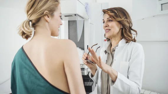
The challenge for clinicians, they note, is that disparate technologies require women to position themselves differently. A woman stands when scanned by x-ray mammography, and if referred for ultrasound, she lies on her back, or her stomach if MRI is used.
But the breast is a soft organ that changes shape in each position, with cancerous lesions moving around, too, becoming more difficult for radiologists to pinpoint. Sometimes, second-look ultrasounds can force providers to spend an hour just to relocate an abnormality.
Their technology would help to merge all of these different images to provide caregivers with a much clearer picture, according to a Dec. 11 announcement from the university.
“We're using biomechanics to predict the movements of those suspicious tissues, so when a person is lying on her back, such as during surgery, the surgeon can be provided with a much better idea as to where to find the suspicious lesion,” Babarenda Gamage, PhD, a biomedical engineer with the University of Auckland and investigator on the project, said in a statement.
Their biomechanical analysis technique will allow clinicians to co-locate signs of cancer, such as micro-calcifications, using both x-rays and MRI. Modeling the breast has proven difficult, they noted, with wide variance in tissue from one woman to the next. The team has been granted access to hundreds of scans from the Auckland District Health Board to aid in that work.
They’re making progress in those modeling methods, and hope this machine learning tool will be put into practice soon.
“This work is approaching real-time clinical application, which is very exciting in terms of realizing the benefits of advanced computational techniques in improving outcomes for patients,” said Anthony Doyle, MD, an MRI expert at Auckland City Hospital, who has added University of Auckland investigators on the project.

Marty Stempniak has covered healthcare since 2012, with his byline appearing in the American Hospital Association's member magazine, Modern Healthcare and McKnight's. Prior to that, he wrote about village government and local business for his hometown newspaper in Oak Park, Illinois. He won a Peter Lisagor and Gold EXCEL awards in 2017 for his coverage of the opioid epidemic.