
Some describe it as an electric, vibrating sensation, as if something were crawling under their skin. To others, the symptoms take the form of persistent headaches, bone and joint pain, tightness of the hands and feet, and a cognitive funk or “brain fog.” For Gena Norris, the wife of actor, martial artist and product pitchman Chuck Norris, the reaction to what she charges is gadolinium-based contrast agent (GBCA) was even more debilitating.
In a $10 million lawsuit filed in November 2017 against manufacturers and distributors of the agent, she cited a burning pain throughout her body coupled with violent shaking, numbness, weakness, kidney damage and trouble breathing. She maintains that these symptoms trace to three contrast-enhanced MRIs she received in a one-week period.
Once considered yesterday’s headlines, GBCAs and their pernicious offspring, gadolinium deposition disease, are back in the news in a way that has radiologists fretting and fidgeting more than usual. Turn on the TV and watch the ads from law firms trolling for class-action clients willing to pursue the possibility that they “may be entitled to compensation.” Or go to the website of patient advocacy groups like the Lighthouse Project and read how one of its founders, Sharon Williams, developed a rash on her lower legs some 18 days after her fifth dose of contrast and now r eports that her brain “feels like it has been cut in half with frequent pain on the left side and non-stop, 24/7 pressure on the right side.”
What to make of these cascading eruptions? Is gadolinium poisoning a bona fide crisis in the making for radiology or another overblown chapter in the ongoing gadolinium saga?
One telling sign of where developments are headed was a gathering in February 2018 of 60 experts from around the world. Working with RSNA and the ACR, the National Institutes of Health organized the workshop, which aimed at bridging the gap between the ongoing public concerns and much prior research showing gadolinium to be safe when used judiciously.
“I think it’s appropriate that we maintain our due diligence and study the potential clinical effects,” acknowledges one of those invited to the summit, Robert McDonald, MD, PhD, a neuroradiologist at the Mayo Clinic.
Studies led or co-led by McDonald have found that traces of gadolinium deposition can remain in brain tissue for years after administration as MRI contrast. This effect bears continued watchfulness, he says, “but we are at risk of causing fear-mongering, which does not benefit us as providers or our patients. I now have patients who are so fearful of getting gadolinium contrast that they’re willing to compromise their health.”
In Search of a Smoking Gun
Gadolinium-based contrast agents—which have been a staple in radiology suites for the past 30 years—are considered to have good safety and benefit-to-risk profiles. The only known disease linkage has been to nephrogenic systemic fibrosis (NSF) in patients with impaired renal function, a phenomenon uncovered in 2006 that has since been neutralized through more careful screening of patients and boxed warnings mandated by the FDA.
As experts in the field hasten to point out, there has never been a prospective study tying GBCAs to any adverse disease pathology.
“There is concern, but what’s seriously missing is reproducible, scientific, sound evidence,” says Emanuel Kanal, MD, professor of radiology and neuroradiology at University of Pittsburgh Medical Center (UPMC), who has extensively studied MRI contrast agents.
Indeed, it’s unclear to many professionals whether gadolinium deposition disease—a name coined several years ago to describe a spectrum of conditions that can arise hours to two months after the administration of a contrast agent—is actually a disease or a set of symptoms in search of a disease.
“I feel for the patients who report these symptoms, but, as a physician and scientist, I have trouble putting them together with gadolinium, given their variety of presentations and the usually immediate reactions that are reported,” says Howard Rowley, MD, a professor of radiology, neurology and neurosurgery at the University of Wisconsin School of Medicine and Public Health. Rowley says he doesn’t consider the occurrence of reported symptoms a signpost of disease but, instead, “an observation at this point. I personally doubt it has to do with gadolinium retention.”
On and Off the Radar
What’s indisputable is the ubiquity of MRIs enhanced with contrast agents, both linear and macrocyclic. Some 450 million injections of contrast agents have been administered worldwide since 1988, and of the 36 million MRs performed in U.S. hospitals in 2017, nearly 40 percent used contrast dyes, according to a recent study, “Retention Concerns About MR Studies Using Gadolinium-Based Contrast Agents” (JACR, June 2018).
In fact, GBCAs have become so dominant, the study points out, that gadolinium concentration in the surface water of San Francisco Bay has increased “by nearly an order of magnitude over the past 20 years,” the authors report, and has also been identified in waterworks along the Ruhr River in Germany.
After the NSF entanglement, GBCAs raised little fuss until 2014, when studies began to show high signal intensity, or hypersensitivity, in brain tissue of patients after repeat administrations of GBCA (mostly linear chelating agents). What’s more, intracranial gadolinium accumulation appeared to be a lifetime cumulative event that could be observed after as few as four administered doses.
These findings caught many radiologists off guard inasmuch as they thought gadolinium deposition couldn’t occur in patients with normal renal function. The terms “gadolinium storage condition” and “gadolinium deposition disease” were born and, quite quickly, became the subjects of dozens of investigations.
Most professionals now accept the fact that GBCAs are associated with some gadolinium retention in organs like the brain, bones and skin, and that macrocyclic agents show significantly lower gadolinium uptake than their linear counterparts, thanks to the former’s cage-like structure.
But the fact that these studies have noticeably failed to remove the cloud hanging over contrast agents only increases the pressure on radiology to continue digging.
Meanwhile, GBCAs have morphed into a barbed Catch-22 for many radiologists, particularly neuroradiologists who typically give contrast to upwards of 45 percent of their patients undergoing MRI exams.
“We can find metastatic disease when it’s of millimeter size in the brain, but it may be impossible to do that without contrast,” explains Kanal, adding that GBCAs “may allow me to detect something when it’s tiny and still treatable.”
McDonald offers further context. “Gadolinium contrast has literally advanced the field of MRI by decades,” he says, “and if we were to take it away we would set the field back 20 to 30 years in terms of our ability to diagnose disease. These are facts which, unfortunately, are not being conveyed to patients.”
 Better Safe Than Sorry?
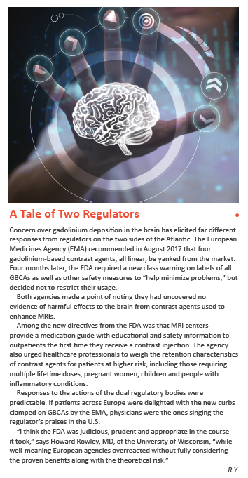
Patient and consumer advocates have not been without their successes, though most would claim they are insufficient. The European Medicines Agency (EMA), for one, recommended that most linear GBCAs be withdrawn from the market, while the FDA in December 2017 issued a new class warning and other safety measures for all gadolinium-based contrast agents in response to the public‘s metastasizing concern. It also urged radiologists to “limit GBCA use to circumstances in which additional information provided by the contrast agent is necessary.” The agency found no evidence of harm to humans, however, that might justify removing or restricting the agents from the market. (See accompanying article “A Tale of Two Regulators.”)
Still, the brouhaha fueled by an increasingly vocal patient constituency has not been lost on imagers.
“What I’m seeing across the board is many hospitals and clinics switching their contrast agents from linear to macrocyclic molecules,” says Mahadevappa Mahesh, MS, PhD, chief physicist and a professor of radiology and cardiology at Johns Hopkins, adding that his own institution became part of that trend over the past year.
Similarly, at UPMC, Kanal reports that he cancels a quarter of the requests for contrast that cross his desk, then openly discusses his decision with the referring physician who originated the request.
Kanal is well positioned to opine on GBCAs and, more precisely, on the conventional wisdom that macrocyclic agents as a whole are safer than linear, which allow more heavy metal to escape flushing through the kidneys and instead take up residence inside human tissue. In reality, as Kanal posited in a recent study intriguingly titled “Gadolinium-based Contrast Agents: The Plot Thickens” (Radiology, November 2017), there are significant differences within each class in terms of degree or rate with which gadolinium is retained among members of the same group.
“Lumping together” all macrocyclic and all linear agents as if they are interchangeable may be convenient, his study points out, but the soundness of such a move is “not supported by reality.” The point is underscored by McDonald, a member of the ACR’s committee on drugs and contrast media. “As macrocyclic agents are taking over the market,” he remarks, “we’re seeing quite a few complaints of gadolinium deposition disease from these agents.” He terms the development puzzling in light of macrocyclics’ presumed lower gadolinium deposition compared to linear.
As mysteries around gadolinium continue to confound, at least one appears to have been resolved: how retained gadolinium could make its way to the side of an otherwise intact blood-brain barrier.
The conjecture by some was that the passage involved irreparable damage to the brain. That appears not to be the case, however. A growing body of evidence suggests that cerebrospinal fluid and the glymphatic system are the real carriers of contrast to the brain, and that the vast amount of deposition in that organ is actually stuck in the capillary walls, not in brain tissue.
No study has yet to find even microscopic signs of damage to the brain structure.
A Question of Balance
Where does all the noise and confusion leave patients who report gadolinium disease-like symptoms? Here’s the good news for this group: If the radiology community once paid mere lip service to people voicing concerns over gadolinium, physicians are now listening carefully.
A rash of studies is underway to determine the biological activity of gadolinium that gets left behind, and growing numbers of radiologists are welcoming questions from MRI patients about the risks of gadolinium retention.
Still, after all the poking and prodding gadolinium has received in recent years, it remains true that more is unknown about the relative toxicity of the substance than is known.
“Until we find evidence of harm, I don’t think we need to modify our practice,” insists McDonald. “But for me personally, [the controversy] has made me more thoughtful about when to give contrast and to whom. Above all, we need to ask ourselves as radiologists: ‘Is this agent going to potentially provide clinically useful and beneficial information for the patient?’”